We’re upgrading our hospital and working to keep disruption to a minimum. Learn more.
Ultrasound Scan
Also known as: ultrasound, sonogram, ultrasonography, diagnostic ultrasound, USG
What is an ultrasound scan?
An ultrasound scan, also known as ultrasound imaging, uses high-frequency sound waves to produce an image of the body’s internal structures. Ultrasound can be used for patients of any age.
Why you might need an ultrasound scan
An ultrasound scan uses high-frequency sound waves to produce detailed images of your body’s interior. This non-invasive method is commonly employed to examine various organs and tissues, including the liver, gallbladder, pancreas, kidneys, and thyroid gland. By using sound waves to create these images, ultrasound scans can help diagnose conditions such as gallstones, kidney stones, and liver disease. Additionally, they are invaluable in guiding biopsies and other medical procedures. One of the significant advantages of ultrasound scans is that they do not use ionizing radiation, making them a safe and effective tool for diagnosing a wide range of medical conditions.
Types of ultrasound scans
There are two types of ultrasound scan: external and internal. An internal ultrasound scan involves the insertion of a probe into the body, providing detailed images of internal organs and tissues. The type of scan you receive depends on the part of your body being scanned and the reason for the scan.
Internal ultrasound scans are used to examine organs such as the prostate gland, ovaries or womb; these can also be scanned using an external ultrasound scan, if requested.
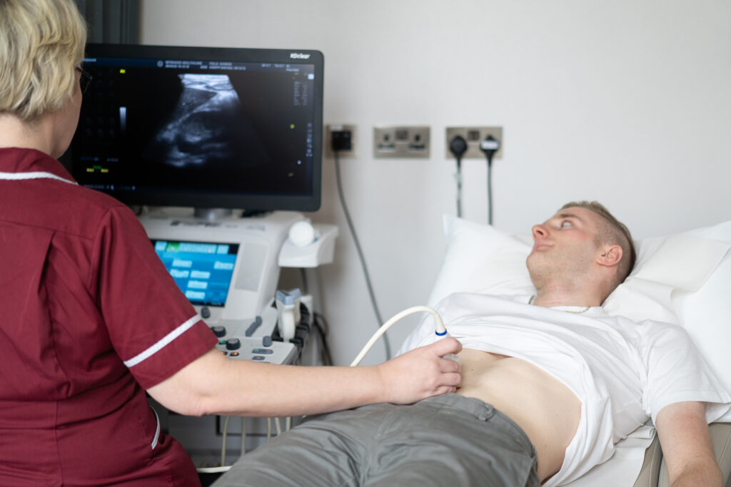
External ultrasound scans are used to examine the heart, liver, kidneys and other abdominal organs. A variety of muscles, joints and tendons can also be scanned this way.
Specialist Ultrasound Techniques
Ultrasound technology has advanced to include several specialist techniques designed to examine specific areas of the body or diagnose particular conditions. These techniques include:
- Doppler Ultrasound: This method uses high-frequency sound waves to measure blood flow and detect abnormalities in blood vessels. It is particularly useful for diagnosing vascular diseases and monitoring blood flow in various organs.
- Transvaginal Ultrasound: In this technique, a probe is inserted into the vagina to provide detailed images of the ovaries, womb, and surrounding structures. It is commonly used in gynaecological examinations.
- Transrectal Ultrasound: This involves inserting a probe into the rectum to examine the prostate gland and surrounding tissues. It is often used in diagnosing prostate conditions and guiding prostate biopsies.
Guide price: from £450*
More about pricingBefore the test
To undergo an ultrasound scan, you’ll need a referral from a healthcare professional. Upon arrival, an imaging staff member will guide you through the procedure. A Consultant Radiologist will conduct the ultrasound scan. Depending on the specific area being examined, you might receive special instructions to enhance image quality. For instance, if you’re having a scan of the digestive system, liver, or gallbladder, you may need to refrain from eating for several hours beforehand. For pelvic and renal scans, it’s important to drink plenty of water to ensure your bladder is full during the scan. Additionally, for certain interventional procedures, you may be asked about your current medications, as some may need to be paused prior to the scan.
It’s also essential to wear comfortable clothing and possibly change into a hospital gown, depending on the area being scanned. The imaging staff will ensure you are comfortable and well-informed about the process, addressing any concerns you might have. If you are undergoing an internal ultrasound, such as a transvaginal or transrectal ultrasound, you will be briefed on what to expect, as these procedures involve inserting a probe into the body to obtain detailed images.
Furthermore, if you have any medical devices, such as an intrauterine device (IUD) or pacemaker, inform the healthcare team beforehand, as this information can be crucial for the examination. For those with allergies, particularly to latex, it’s important to notify the staff to prevent any possible reactions during the procedure.
During the test
Most ultrasound scans take between 15 and 30 minutes to complete, depending on the specific type of scan being conducted. However, the duration can vary based on the complexity of the examination and the area being scanned. For instance, a comprehensive abdominal ultrasound might take longer than a focused scan of a smaller region.
Ultrasound scans are considered safe, with no known risks associated with their use. They do not involve exposure to ionizing radiation, which is a significant advantage over other imaging tests like X-rays and CT scans. This makes ultrasounds particularly suitable for pregnant women and children. The safety of ultrasound scans is supported by extensive research and the endorsement of medical bodies such as the British Medical Ultrasound Society.
During an internal ultrasound, a small, sterile ultrasound probe is carefully inserted into the vagina or rectum. This procedure may cause slight discomfort but should not be painful. The healthcare professional conducting the scan will explain each step, ensuring the patient’s comfort and addressing any concerns. Internal ultrasounds provide detailed images of internal structures, which are crucial for diagnosing conditions affecting the pelvic organs, prostate gland, and other areas. These scans are invaluable for evaluating blood flow and detecting abnormalities such as tumours, cysts, or inflammation.
The ultrasound probe, also referred to as a transducer, is a handheld device that is integral to the ultrasound scanning process. When placed on the body’s surface or inserted internally, the probe emits sound waves that reflect off internal structures. These reflected sound waves are captured by the probe and converted into electrical signals. The ultrasound machine processes these signals to generate real-time images displayed on a monitor. The clarity of these images is vital for accurate diagnosis, making the ultrasound probe an essential component of the ultrasound machine.
After the test
After completing your ultrasound scan, the images will be evaluated by a radiologist or healthcare provider who will assess them for any abnormalities related to the reason for your scan. They will interpret the results and prepare a report, which may take some time, depending on the complexity. Once the results are ready, your healthcare provider will go over the findings with you. If anything unusual is detected, they may recommend further testing or treatment. If everything looks normal, you might just be advised to follow up as needed. Since ultrasound scans are non-invasive, there’s no recovery time, and you can resume your normal activities right away.
Appointment and Treatment Plan
Referral
You will first need a referral from your doctor to schedule an appointment with the imaging department for your ultrasound.
Preparation
Before the examination, the technician will ask you to remove any clothing from the area being scanned. For an external ultrasound, they may ask you to wear a gown or remove clothing from the waist up. If it’s an internal ultrasound (like a pelvic or transvaginal ultrasound), the technician may provide you with a gown and explain any additional preparation instructions, such as needing a full bladder for certain scans.
Positioning
For an external ultrasound, you will typically lie down on an examination table. The technician will apply a gel to the area being scanned, which helps with the transmission of sound waves. They will then move the transducer (a small handheld device) over your skin to capture images of the internal organs or tissues.
For an internal ultrasound, the technician will guide a special probe into the body (like a transvaginal probe for pelvic scans). This allows for a closer view of the internal organs, such as the uterus or ovaries.
Image Production
The technician uses the transducer to capture detailed images. These images are then sent to a radiologist for review.
Image Interpretation
The radiologist will interpret the ultrasound images, looking for any abnormalities or conditions that may require further investigation or treatment. This step is crucial for diagnosing conditions such as cysts, tumours, or inflammation, and for planning any necessary follow-up care.
Follow Up
If any abnormalities have been detected during the scan, you will attend a follow-up appointment with a consultant to discuss next steps relating to your treatment plan.
Experts
We are proud to provide patients with access to a wide range of clinicians, chosen specifically for their knowledge and reputation in their area of expertise. Our experts align with our values: putting you at the centre of your care and educating you on your options at each step of the journey. We encourage you to learn more about our clinicians and how they can help you below. As always, please contact our patient services team if you require any additional information.
We offer 3 ways to pay for your treatment
We exist to take the stress out of private healthcare.
Our payment options are designed to offer you easy access to our treatments and services. You can choose to pay on the day, spread the cost, or use your private medical insurance.
Our patient services team will guide you through the process, providing clear costs and support throughout your course of treatment so you can focus on the thing that matters most – your health.
Whether you pay in advance, spread the cost, or use your private medical insurance, rest assured you will be receiving exceptional care 365 days a year.
Pay in Advance
Even if you do not have medical insurance, you can still get quick and comprehensive access to private medical care.
We provide transparent pricing from your initial consultation to the completion of your treatment so you know where you stand, every step of the way.
We accept all major debit and credit cards, as well as Apple Pay for UK residents. Please note that we do not accept cash or cheques.
Spread the cost monthly
Paying for your treatment at One Stop Healthcare can be spread monthly from 12 to 60 months, rather than paying in one go.
With an upfront 10% deposit paid, via our Financial partner Chrysalis Finance, we offer various flexible terms to enable you to spread the cost, including 12-months at 0% APR. Click here to find out more.
Monthly payments need to be linked to a One Stop Healthcare treatment over £385 and is subject to a 14-day ‘cooling-off’ period before any treatment can start.
Your on-going payments will be made directly between Chrysalis and yourself. It’s that simple.
Pay using PMI
We are recognised by all major health insurance companies and with our extensive range of services, there are lots of benefits to using your insurance with us. Our patient services team is here to answer any questions you may have about using your private health insurance with us.
Please bring along your policy details including your scheme details, membership or policy number, expiry date and confirmation of eligibility to claim (i.e. your authorisation number). If you do not have these details with you, we will require payment from you on the day. Patients are liable for any amounts not settled by their insurer.
Pricing
Price depends on areas scanned.
| Ultrasound 1 part: | £450 |
| Ultrasound 2 part: | £490 |
| Transvaginal and Abdomen/Pelvis: | £520 |
| Ultrasound Testes: | £420 |
| Breast Unilateral: | £450 |
| Breast Bilateral: | £520 |
| Ultrasound Echo: | £575 |
| Ultrasound Guided Injection 1 part: | £700 |
| PRP 1 Injection: | £1,100 |
| PRP 1 Injection (with HA): | £1,200 |
| PRP 3 Injections (with HA): | £2,900 |