We’re upgrading our hospital and working to keep disruption to a minimum. Learn more.
MRI Scan
Also known as: Magnetic Resonance Imaging
An MRI is a medical scan that captures detailed images of the body’s internal structures. It’s widely used to diagnose and monitor various conditions without using harmful radiation.
What is Magnetic Resonance Imaging (MRI)?
MRI (Magnetic Resonance Imaging) is an advanced diagnostic technique that utilises strong magnets and radio waves to generate detailed images of the body’s internal structures. Unlike traditional imaging methods such as X-ray imaging, which utilise ionising radiation, MRI is a non-invasive and safer alternative. MRI generates high-resolution images of anatomy and physiological processes, enabling doctors to see internal structures not visible on X-rays.
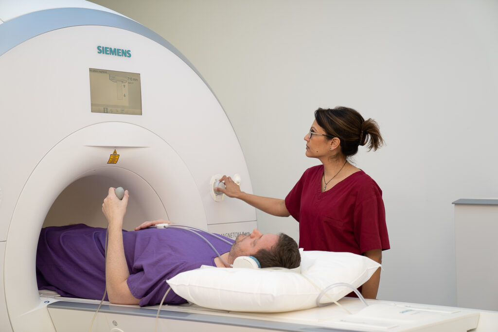
The process of MRI scanning typically involves a patient lying on a table that slides into a large tube-like MRI machine. This machine houses the magnets and radiofrequency systems necessary to create the detailed images. MRI scans diagnose various conditions, such as brain disorders, joint injuries, and soft tissue abnormalities. In particular, MRI scanners are invaluable for detecting injuries, tumours, and heart problems.
A significant advantage of MRI over CT and PET scans is its lack of ionising radiation, making it safer for frequent imaging. This is particularly important for patients requiring multiple scans over time, as it reduces the risk of radiation exposure. Moreover, MRI does not involve surgical procedures, highlighting its non-invasive nature.
How does an MRI scan work?
MRI (Magnetic Resonance Imaging) allows doctors to view detailed internal body images without radiation. It works by using a strong magnet to create a magnetic field, which affects tiny hydrogen atoms found in the water and fat inside your body.
During the scan, the machine sends gentle radio waves that temporarily move these hydrogen atoms out of alignment. When the waves stop, the atoms return to their normal position and release signals. The MRI scanner detects these signals, and a computer processes them to create detailed images of your organs, muscles, and other soft tissues.
Different types of tissues give off signals in slightly different ways, which helps the MRI distinguish between them. This difference creates contrast in the images, allowing doctors to see even small details that might indicate a health condition. By carefully balancing magnetic fields, radio waves, and tissue characteristics, MRI provides clear, high-resolution images that help with accurate diagnosis and treatment.
Types of MRI scans
MRI technology is versatile, offering various scans tailored to specific medical needs. Among the most prominent are Cardiac MRI and Breast MRI. Each MRI type serves a unique purpose, from assessing brain activity to evaluating heart conditions and detecting breast cancer.
Cardiac MRI
Cardiac MRI is a specialised type of MRI scan designed to evaluate heart and blood vessel conditions. It provides detailed images of the heart’s structure, function, and blood flow, allowing doctors to assess the heart’s pumping efficiency and the condition of its tissues. This type of MRI is particularly useful for diagnosing conditions such as heart disease, congenital heart defects, and cardiomyopathies.
One of the key applications of cardiac magnetic resonance imaging is Magnetic Resonance Angiography (MRA), which generates detailed images of the arteries. MRA is instrumental in evaluating stenosis (narrowing of the arteries) or aneurysms (abnormal bulging of the artery walls). By providing comprehensive images of the cardiovascular system, cardiac MRI helps in planning treatments and monitoring the effectiveness of interventions.
Breast MRI
Breast MRI is particularly beneficial for patients with dense breast tissue. Dense tissue can make it difficult to detect abnormalities using traditional imaging techniques like mammography. Breast MRI uses strong magnets and radio waves to create detailed images of the breast, making it easier to identify any unusual changes.
This type of MRI is especially useful for detecting breast cancer, particularly in individuals with high-risk factors or a family history of the disease. By providing high-resolution images of the breast tissue, Breast MRI can help in early detection, diagnosis, and treatment planning, offering a critical advantage in the fight against breast cancer.
Early detection saves lives. Our dedicated MRI Breast imaging offers unmatched clarity for high-risk screening, dense breast tissue evaluation, and follow-up care. With a focus on patient comfort and clinical accuracy, we empower you and your care team with detailed insights for informed decisions.
Prostate MRI
Take charge of your prostate health with our advanced MRI Prostate imaging. Using multi-parametric MRI (mpMRI), we provide high-resolution, non-invasive scans that support accurate diagnosis and targeted treatment planning. Ideal for early cancer detection and ongoing monitoring, our prostate MRI delivers peace of mind through precision.
MSK (Musculoskeletal) MRI
Don’t let joint or muscle pain hold you back. Our MRI MSK scans deliver crystal-clear images of bones, ligaments, tendons, and soft tissues, helping diagnose injuries and chronic conditions with confidence. Whether you’re an athlete or managing daily discomfort, we help get you moving again.
Liver MRI
Your liver plays a vital role – keep it in check with advanced MRI Liver imaging. Our scans provide detailed assessment of liver lesions, fibrosis, fatty liver disease and more, often without the need for contrast. Fast, accurate and non-invasive, delivered by experienced specialists you can trust.
Safety and risks of MRI Scans
While MRI scans are generally considered safe, it is essential to follow specific guidelines to minimise any risks. One of the significant safety advantages of MRI over other imaging techniques like CT scans and PET scans is that MRI does not use ionising radiation. This makes MRI a safer option, particularly for patients who require frequent imaging.
However, there are some precautions to consider. Patients with metal objects in or on their bodies may need extra precautions during MRI scans. Metal implants, such as screws, rods, stents, or surgical clips, do not automatically disqualify a patient from undergoing an MRI, but it is crucial to inform the medical staff to assess potential risks. Some medical devices, like pacemakers or implantable cardioverter-defibrillators, may be made MRI-safe or monitored during the procedure.
Radiographers play a vital role in ensuring that MRI scans are conducted safely, especially for patients with known metal implants. They follow strict protocols to minimise any risks and ensure a safe and comfortable experience for the patient. By adhering to these safety guidelines, MRI remains a reliable and safe diagnostic tool.
Guide price: from £580*
More about pricingBefore the scan
To undergo an MRI scan, you must have a referral from a doctor or a registered healthcare practitioner such as a physiotherapist, osteopath or chiropractor.
Preparing for an MRI scan involves several important steps to ensure a smooth and successful procedure. You are advised to wear comfortable clothing that is easy to remove, as you will likely need to change into a hospital gown. It is crucial to avoid wearing jewellery or any metal items to prevent interference with the MRI machine. If you are having a scan of your abdomen and pelvis you should not have anything to eat or drink for 2 hours before your appointment. Small sips of water are acceptable. If you’re having an MRI of your head or orbits (eye sockets), you should not wear any eye make-up when you attend your appointment.
Before the scan, make sure to inform the MRI technician about any metal in your body, including implants or medical devices. You will typically be required to fill out a safety checklist before your MRI appointment. This checklist helps the medical staff assess any potential risks and ensure your safety during the scan.
If you might experience claustrophobia, it is advisable to contact the scanning department before your appointment to discuss any concerns. Some MRI machines are designed to be less confined, which can help alleviate feelings of discomfort. If necessary, you can discuss sedation options with your doctor to make the experience more manageable. It is also recommended to eat normally before the MRI unless advised otherwise for specific scans.
Communication with the MRI technologist is essential throughout the procedure. Arrive on time for your MRI appointment to ensure a smooth process. If you feel uncomfortable or experience a panic attack during the scan, you can use the call button provided to alert the technologist. By following these preparation steps, you can help ensure a successful and stress-free MRI experience.
During the scan
During an MRI scan, you will lie on a table that slides into a large tube-like MRI machine. The entire process may take between 15 to 90 minutes, depending on the area being examined. Remaining still during the scan is essential for clear images. Some MRI scans may involve using a frame over the area being scanned, which helps improve image quality.
One of the most noticeable aspects of an MRI scan is the loud tapping sounds caused by the switching of electric current in the scanner coils. To help manage the noise, you are often provided with headphones or earplugs. The MRI technician will communicate with you through an intercom, ensuring comfort and information throughout the procedure.
A family member or friend may be allowed to accompany you during the MRI scan for additional support. This can be particularly helpful if you feel anxious or claustrophobic. By understanding what to expect during the MRI scan, you can be better prepared and more at ease during the procedure.
MRI with contrast
MRI with contrast involves the use of contrast agents to enhance the clarity of the images. These agents help in visualising specific tissues and blood vessels more distinctly, making certain details clearer in the MRI exam. The contrast material is typically administered through an intravenous catheter inserted into a vein.
Common side effects from contrast agents include nausea, skin rashes, and headaches. There is a very slight risk of allergic reactions to the contrast material, which are usually mild and controllable. Symptoms of an allergic reaction may include feeling weak, sweating, and difficulty breathing. It is important to disclose any history of allergic reactions or blood clotting issues before receiving a contrast agent.
If you have severe kidney issues, you may face risks from contrast agents and may require a kidney function test before administration. If you start to feel unwell after the contrast injection during an MRI, inform the radiographer immediately. Swelling or pain at the injection site should also be reported to the radiographer and your GP if it worsens.
After the scan
After the MRI scan, a radiologist will analyse the images and will usually provide a report to your referring healthcare provider within 48 hours, but some specialist scans may take longer. The report includes detailed findings from the MRI images, essential for diagnosing complex conditions and planning treatments. The radiologist primarily communicates these findings through written reports directed to your referring physician.
Typically, it can take 1 to 2 weeks to receive MRI results. If you have not received your MRI results after this period, you should contact the doctor who arranged the test.
Your referring healthcare provider will discuss the results with you and determine the next steps in your treatment plan. By understanding the post-scan process, you can be better prepared for what to expect after your MRI scan.
Appointment and Treatment Plan
Referral
To begin, you will need a referral from a doctor or a registered healthcare practitioner such as a physiotherapist, osteopath or chiropractor.
Preparation
Preparing for an MRI scan involves several important steps to ensure a smooth and successful procedure. You are advised to wear comfortable clothing that is easy to remove, as you will likely need to change into a hospital gown. It is crucial to avoid wearing jewellery or any metal items to prevent interference with the MRI machine. Depending on the type of scan performed, you might need to follow additional preparation steps.
Positioning
After the MRI scan, a radiologist will analyse the images and provide a report to your referring healthcare provider within 48 hours. The report includes detailed findings from the MRI images, essential for diagnosing complex conditions and planning treatments.
Follow-up
You will need to book a follow-up appointment with your referring healthcare provider. Based on the findings, they may recommend further tests or treatments.
Experts
We are proud to provide patients with access to a wide range of clinicians, chosen specifically for their knowledge and reputation in their area of expertise. Our experts align with our values: putting you at the centre of your care and educating you on your options at each step of the journey. We encourage you to learn more about our clinicians and how they can help you below. As always, please contact our patient services team if you require any additional information.
We offer 3 ways to pay for your treatment
We exist to take the stress out of private healthcare.
Our payment options are designed to offer you easy access to our treatments and services. You can choose to pay on the day, spread the cost, or use your private medical insurance.
Our patient services team will guide you through the process, providing clear costs and support throughout your course of treatment so you can focus on the thing that matters most – your health.
Whether you pay in advance, spread the cost, or use your private medical insurance, rest assured you will be receiving exceptional care 365 days a year.
Pay in Advance
Even if you do not have medical insurance, you can still get quick and comprehensive access to private medical care.
We provide transparent pricing from your initial consultation to the completion of your treatment so you know where you stand, every step of the way.
We accept all major debit and credit cards, as well as Apple Pay for UK residents. Please note that we do not accept cash or cheques.
Spread the cost monthly
Paying for your treatment at One Stop Healthcare can be spread monthly from 12 to 60 months, rather than paying in one go.
With an upfront 10% deposit paid, via our Financial partner Chrysalis Finance, we offer various flexible terms to enable you to spread the cost, including 12-months at 0% APR. Click here to find out more.
Monthly payments need to be linked to a One Stop Healthcare treatment over £385 and is subject to a 14-day ‘cooling-off’ period before any treatment can start.
Your on-going payments will be made directly between Chrysalis and yourself. It’s that simple.
Pay using PMI
We are recognised by all major health insurance companies and with our extensive range of services, there are lots of benefits to using your insurance with us. Our patient services team is here to answer any questions you may have about using your private health insurance with us.
Please bring along your policy details including your scheme details, membership or policy number, expiry date and confirmation of eligibility to claim (i.e. your authorisation number). If you do not have these details with you, we will require payment from you on the day. Patients are liable for any amounts not settled by their insurer.
Pricing
Scan price subject to bespoke quotation
*Additional consultant fee not included
| 1 part: | £580 |
| 2 part: | £940 |
| 3 part: | £1,300 |
| 4 part: | £1,400 |
| 5 part: | £1,600 |
| 6 part: | £1,900 |
| Arthrogram: | £1,700 |
| Small Bowel (including contrast) | £1,300 |
| Contrast Agent: | £130 |
| Primovast Contrast: | £160 |
| MRI Cardiac: | On request |
FAQs
MRI is generally safe for most individuals, but precautions are crucial for those with metal implants or medical devices. Always inform your MRI technician about any metal in your body to evaluate potential risks.
To prepare for an MRI scan, wear comfortable clothing without metal fasteners, avoid jewellery, and inform the technician about any implants or medical devices. It is also essential to complete a safety checklist prior to your appointment.
During an MRI scan, you can expect to lie on a table that moves into a tube-shaped machine for 15 to 90 minutes, during which you will hear loud tapping sounds. Rest assured; you will maintain communication with the MRI technician throughout the procedure.
Potential side effects of MRI with contrast include nausea, skin rashes, and headaches, along with a slight risk of allergic reactions. Patients with severe kidney issues should have their kidney function assessed prior to the procedure.
MRI results usually take 1 to 2 weeks to be available. If you have not received them by then, it is advisable to reach out to your healthcare provider.