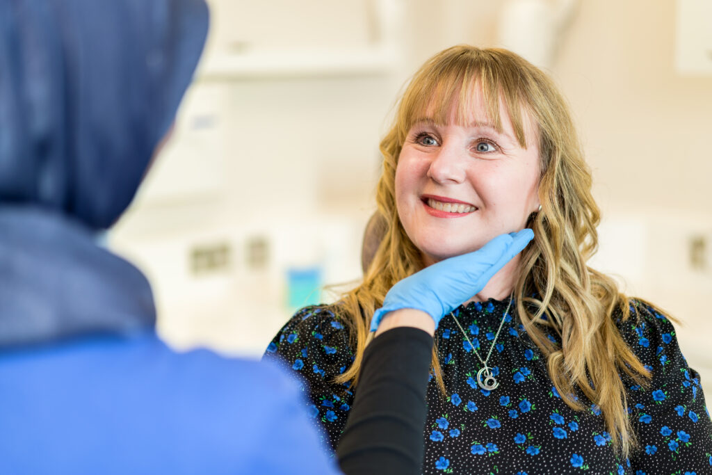
Ear Lesion Removal
Also known as: ear lump removal, surgery to remove a growth on the ear
Excision of an ear lesion is a medical procedure that involves the removal of abnormal tissue from the ear.
Understanding ear lesions
Ear lesions refer to abnormal growths or changes in the skin or tissue of the ear. These lesions can be benign (non-cancerous) or malignant (cancerous). Benign ear lesions are common and can be caused by various factors, including direct and prolonged pressure, poor blood supply, or skin conditions. Malignant ear lesions, on the other hand, are less common and can be caused by skin cancer or other types of cancer.
Ear lesions can affect any part of the ear, including the outer ear, ear canal, middle ear, and inner ear. It is important to seek medical attention if you notice any unusual changes in the skin of your ear. Swelling around the ear can also be a sign of an underlying ear lesion.
Early diagnosis and treatment are crucial for preventing complications and ensuring the best possible outcome.
Diagnosis and treatment
Diagnosing ear lesions typically involves a thorough physical examination, a review of medical history, and imaging tests such as X-rays or CT scans. In some cases, a biopsy may be performed to determine if the lesion is benign or malignant.
Treatment for ear lesions depends on the type, size, and location of the lesion. Benign ear lesions can often be treated with topical creams, liquid nitrogen, or surgical excision. Malignant ear lesions, however, may require more aggressive treatments such as surgery, radiation therapy, or chemotherapy. Minimally invasive procedures like radiosurgery or laser therapy can also be effective in some instances. Consulting a specialist is crucial to determine the most appropriate treatment plan for ear lesions.
Complications and risks
If left untreated, ear lesions can lead to a range of complications. Lesions that affect the middle or inner ear can result in hearing loss, which significantly impacts communication and quality of life. Malignant lesions, in particular, may cause persistent discomfort and pain, adding to the patient’s distress. Infections can also develop if lesions become irritated or ulcerated, potentially leading to more serious health concerns. Prompt medical evaluation and treatment are essential to minimise these risks and ensure appropriate management.
Reconstruction and rehabilitation
In some cases, treating ear lesions may require reconstructive or rehabilitative efforts to restore both form and function. Skin grafting may be used to cover areas affected by surgical excision, especially when a significant amount of tissue has been removed. In instances of severe damage or disfigurement, prosthetic ears can help restore appearance. If the lesion has compromised hearing, hearing aids may be recommended to support auditory function. Physical therapy may also play a role, particularly when mobility or balance is affected. These interventions can significantly improve both the appearance and functionality of the ear, contributing to a better overall quality of life.
Please call to enquire about the price
Ways to payBefore surgery
Initial consultation
The initial consultation is a crucial step in the process of removing ear lesions. During this appointment, the doctor will conduct a comprehensive examination of the affected ear, paying close attention to the size, shape, colour, and location of the lesion. The doctor may inquire about the duration of the lesion, any associated symptoms such as pain or discharge, and any history of skin cancer or other relevant medical conditions.
In some cases, the doctor may perform a biopsy during the consultation to obtain a small tissue sample from the lesion. This helps in accurately diagnosing the lesion type, distinguishing between benign and malignant growths, and planning appropriate treatment.
The consultation also provides an opportunity for you to discuss your concerns, understand the procedure options, and ask questions about the risks, benefits, and expected outcomes of the ear lesion removal. The doctor will explain the recommended treatment approach, which may involve surgical excision, cryotherapy, or laser removal, and outline what you can expect before, during, and after the procedure.
Additionally, the doctor will review your medical history, including any medications, allergies, or underlying health issues that may affect the surgery or recovery. You may be advised to stop certain medications, such as blood thinners, before surgery to minimise bleeding risks.
Preparing for surgery
Before undergoing ear surgery, it is crucial to follow specific preparation guidelines to ensure the best possible outcome. It is often recommended to avoid certain medications, such as blood thinners, in the days leading up to the surgery.
You will also need to arrange for transportation to and from the hospital, as you will not be able to drive yourself after the procedure. Fasting for a specified period before the surgery is typically required, and you should follow your surgeon’s instructions regarding food and drink intake.
During surgery
During the procedure, the surgeon will use precise techniques to ensure the best outcome. The external ear is thoroughly cleaned and prepared for the surgical excision.
Local anaesthetic injection is typically administered to numb the affected area, ensuring you experience minimal discomfort throughout the procedure.
The excision involves removing the lesion along with a small margin of healthy tissue to ensure complete removal, especially if cancer cells are suspected to be present. The tissue removed is often sent for biopsy to confirm the diagnosis and rule out malignancy.
In cases where the lesion affects middle-aged or older adults, additional considerations are made to accommodate skin fragility and slower healing times. The surgeon may also take steps to relieve pressure on the tender area during and after surgery to promote healing.
If necessary, neck surgery or further procedures may be discussed if the lesion has spread or if lymph nodes require examination.
Throughout the surgery, the consultant monitors for any signs of complications and ensures that the procedure is completed efficiently, typically allowing patients to go home the same day.
After excision, the wound is carefully closed, often with delicate stitches, and a dressing is applied to protect the area.
The surgeon will provide detailed instructions on wound care, signs of infection, and when to seek medical advice. This comprehensive approach helps ensure optimal healing and reduces the risk of recurrence or complications.
After surgery
Immediate post-surgery care
After undergoing ear surgery, it is crucial to follow your doctor’s post-operative care instructions to ensure a smooth recovery. Swelling around the surgical site is common and should subside within a few days. Keep the area clean and dry, and refrain from any strenuous activities that could impede the healing process.
In the days following the surgery, you may experience some discomfort and mild pain, which can be managed with prescribed medications. Any discharge from the ear should be reported to your doctor immediately. It is also essential to attend all follow-up appointments to monitor your progress and address any concerns that may arise.
Long-term recovery
Long-term recovery after ear lesion removal involves several essential aspects to ensure complete healing and prevent recurrence. You should continue to protect the affected ear from direct and prolonged pressure, as this can contribute to the development of new lesions or delay healing. Wearing a warm hat in cold weather helps maintain good blood circulation to the area, which is crucial for tissue repair.
It is common to experience some residual tenderness or a tender lump at the site where the lesion was removed. This tender area may persist for several weeks but should gradually improve over time. If discomfort occurs or the nodule causes significant pain, you should consult your doctor for further evaluation.
Maintaining good hygiene and avoiding exposure to irritants or excessive moisture can reduce the risk of infection during the recovery period. Patients are advised to avoid inserting objects into the ear canal or applying unprescribed topical agents.
In cases where chondrodermatitis nodularis is diagnosed, additional measures may be necessary to relieve pressure on the affected ear, such as using specialised cushions or avoiding sleeping on the affected side. The British Association for Dermatologists provides detailed leaflets and web links with guidance on managing this condition effectively.
Follow-up appointments are essential for monitoring healing progress and checking for any signs of recurrence or complications. If new symptoms arise, such as swelling, pain, discharge, or changes in hearing, prompt medical attention is recommended.
Overall, long-term recovery emphasises careful care, protection from excessive exposure or pressure, and ongoing communication with healthcare providers to ensure the best possible outcome after ear lesion removal.
Appointment and Treatment Plan
Initial Consultation
Your doctor examines the lesion, reviews your history, and may take a biopsy. You’ll discuss treatment options (surgical, laser, cryotherapy) and get a plan.
Pre-operative Preparation
You may need to stop blood thinners and follow fasting instructions. Arrange transport, and tell your doctor about any meds or health conditions.
Surgery
Performed under local anaesthetic. The lesion is removed with a margin of healthy tissue and the area is closed with stitches. Tissue may be sent for biopsy.
Immediate Post-surgery Care
Expect mild swelling and discomfort. Keep the area clean and dry. Report discharge or signs of infection and attend all follow-ups.
Long-term Recovery
Protect the ear from pressure and cold. Some tenderness may last for weeks. Avoid irritation and consult your doctor if symptoms return. Ongoing care helps prevent recurrence.
Experts
We are proud to provide patients with access to a wide range of clinicians, chosen specifically for their knowledge and reputation in their area of expertise. Our experts align with our values: putting you at the centre of your care and educating you on your options at each step of the journey. We encourage you to learn more about our clinicians and how they can help you below. As always, please contact our patient services team if you require any additional information.
We offer 3 ways to pay for your treatment
We exist to take the stress out of private healthcare.
Our payment options are designed to offer you easy access to our treatments and services. You can choose to pay on the day, spread the cost, or use your private medical insurance.
Our patient services team will guide you through the process, providing clear costs and support throughout your course of treatment so you can focus on the thing that matters most – your health.
Whether you pay in advance, spread the cost, or use your private medical insurance, rest assured you will be receiving exceptional care 365 days a year.
Pay in Advance
Even if you do not have medical insurance, you can still get quick and comprehensive access to private medical care.
We provide transparent pricing from your initial consultation to the completion of your treatment so you know where you stand, every step of the way.
We accept all major debit and credit cards, as well as Apple Pay for UK residents. Please note that we do not accept cash or cheques.
Spread the cost monthly
Paying for your treatment at One Stop Healthcare can be spread monthly from 12 to 60 months, rather than paying in one go.
With an upfront 10% deposit paid, via our Financial partner Chrysalis Finance, we offer various flexible terms to enable you to spread the cost, including 12-months at 0% APR. Click here to find out more.
Monthly payments need to be linked to a One Stop Healthcare treatment over £385 and is subject to a 14-day ‘cooling-off’ period before any treatment can start.
Your on-going payments will be made directly between Chrysalis and yourself. It’s that simple.
Pay using PMI
We are recognised by all major health insurance companies and with our extensive range of services, there are lots of benefits to using your insurance with us. Our patient services team is here to answer any questions you may have about using your private health insurance with us.
Please bring along your policy details including your scheme details, membership or policy number, expiry date and confirmation of eligibility to claim (i.e. your authorisation number). If you do not have these details with you, we will require payment from you on the day. Patients are liable for any amounts not settled by their insurer.
FAQs
If your symptoms return, it is essential to contact your healthcare provider promptly. They may recommend additional treatments or adjustments to your current regimen.
Treatment for an ear lesion depends on the type, size, and cause of the lesion. Standard treatment options include surgical excision, where the lesion is carefully removed under local anaesthetic; cryotherapy, which involves freezing the lesion with liquid nitrogen to destroy abnormal tissue; and laser therapy, which uses focused light to remove or reduce the lesion. In cases of benign lesions, topical medications or observation may be sufficient. For malignant or precancerous lesions, more extensive surgical removal and follow-up care may be necessary to ensure all cancer cells are eliminated. It is essential to consult a doctor for an accurate diagnosis and to determine the most appropriate treatment plan.
The healing time after ear lesion removal varies depending on the size and location of the lesion, as well as the treatment method employed. Typically, small lesions treated with surgical excision or cryotherapy heal within a few weeks. During this time, patients may experience some tenderness, swelling, or mild discomfort in the affected area. Proper wound care, avoiding direct and prolonged pressure, and protecting the ear from cold exposure can promote faster healing. For larger or more complex lesions, especially those requiring reconstruction or skin grafting, healing may take longer. Follow-up appointments are crucial for monitoring the healing process and addressing any complications promptly.
A precancerous lesion on the ear is an abnormal area of skin that has the potential to develop into skin cancer if left untreated. These lesions often result from prolonged exposure to ultraviolet (UV) radiation from the sun or other sources. Examples include actinic keratosis, which appears as rough, scaly patches on the outer ear, and Bowen’s disease, a form of early squamous cell carcinoma in situ. Precancerous lesions may cause symptoms such as redness, scaling, or tenderness, and they require medical evaluation. Early diagnosis and treatment, often involving excision or topical therapies, can prevent progression to invasive cancer and preserve ear function and appearance.
Medically reviewed by Mr Pranay Singh - Consultant ENT Surgeon on 29/07/2025