Carotid Artery Scan
Also known as: neck artery screening, carotid ultrasound, carotid doppler, carotid doppler ultrasound, carotid duplex scan, carotid duplex ultrasonography
A carotid ultrasound is a painless scan that checks the neck arteries for narrowing or blockages that could increase your risk of stroke.
What is the carotid artery?
The carotid arteries are major blood vessels located on each side of your neck. They are responsible for carrying oxygen-rich blood from the heart to the brain and other parts of the head. Each side of the neck contains two carotid arteries: the common carotid artery, which then divides into the internal carotid artery and the external carotid artery. The internal carotid artery supplies blood to the brain, while the external carotid artery supplies blood to the face and scalp. These arteries play a crucial role in maintaining proper blood flow and adequate oxygen supply to the brain, which is vital for normal brain function.
When would a carotid ultrasound be needed?
A carotid ultrasound, also called a carotid artery scan or carotid doppler, is typically recommended if there is concern about carotid artery disease or stenosis, conditions in which the carotid arteries become narrowed or blocked due to plaque buildup. This narrowing can reduce blood flow to the brain and increase the risk of stroke. Your doctor may order this ultrasound scan if you have symptoms such as transient ischemic attacks (TIAs), also known as mini-strokes, or other signs of stroke risk. It may also be advised if you have risk factors like high blood pressure, diabetes, high cholesterol, smoking, or a family history of heart disease or stroke.
Please call to enquire about the price
Ways to payBefore the scan
No special preparation is usually required before a carotid ultrasound. You can eat, drink, and take medications as usual. It is advisable to wear comfortable clothing that allows easy access to the side of your neck. Removing any jewellery or accessories around your neck will help facilitate the scan. Inform your doctor about any medications you are taking and any symptoms you are experiencing.
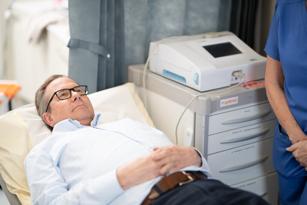
During the scan
During the procedure, you will lie on your back with your head slightly tilted to provide the technician with better access to the carotid arteries on the side of your neck. A clear gel will be applied to your skin to help transmit the ultrasound probe’s sound waves. The technician will gently move the probe along your neck to capture images of the carotid arteries and assess blood flow using Doppler ultrasound technology. This painless procedure usually takes about 15 to 30 minutes and does not involve radiation or needles.
After the scan
After the carotid ultrasound, the images and blood flow data will be analysed by a radiologist, vascular specialist or neurologist. Your doctor will discuss the results with you, explaining whether any narrowing or blockage was detected and what it means for your stroke risk. If plaque buildup or significant narrowing is found, your healthcare provider may recommend lifestyle changes, medications to prevent blood clots or lower cholesterol, or further imaging tests. In severe cases, surgical procedures such as carotid endarterectomy or angioplasty may be considered to restore proper blood flow.
Appointment and Treatment Plan
Getting Ready
No special preparation is needed – you can eat, drink, and take medications as normal. Wear comfortable clothing and remove jewellery around your neck. Let your doctor know about any symptoms or medications you’re taking.
Having the Scan
Lying on your back with your head turned slightly, gel is applied to your neck and the technician moves an ultrasound probe over the carotid arteries to capture images and assess blood flow. The scan is painless, needle-free, and takes around 15-30 minutes.
Understanding Your Results
A specialist reviews the images, and your doctor explains whether any narrowing or blockage was found. Depending on the findings, recommendations may include lifestyle changes, medication, further imaging, or procedures such as carotid endarterectomy or angioplasty if needed.
Experts
We are proud to provide patients with access to a wide range of clinicians, chosen specifically for their knowledge and reputation in their area of expertise. Our experts align with our values: putting you at the centre of your care and educating you on your options at each step of the journey. We encourage you to learn more about our clinicians and how they can help you below. As always, please contact our patient services team if you require any additional information.


We offer 3 ways to pay for your treatment
We exist to take the stress out of private healthcare.
Our payment options are designed to offer you easy access to our treatments and services. You can choose to pay on the day, spread the cost, or use your private medical insurance.
Our patient services team will guide you through the process, providing clear costs and support throughout your course of treatment so you can focus on the thing that matters most – your health.
Whether you pay in advance, spread the cost, or use your private medical insurance, rest assured you will be receiving exceptional care 365 days a year.
Pay in Advance
Even if you do not have medical insurance, you can still get quick and comprehensive access to private medical care.
We provide transparent pricing from your initial consultation to the completion of your treatment so you know where you stand, every step of the way.
We accept all major debit and credit cards, as well as Apple Pay for UK residents. Please note that we do not accept cash or cheques.
Spread the cost monthly
Paying for your treatment at One Stop Healthcare can be spread monthly from 12 to 60 months, rather than paying in one go.
With an upfront 10% deposit paid, via our Financial partner Chrysalis Finance, we offer various flexible terms to enable you to spread the cost, including 12-months at 0% APR. Click here to find out more.
Monthly payments need to be linked to a One Stop Healthcare treatment over £385 and is subject to a 14-day ‘cooling-off’ period before any treatment can start.
Your on-going payments will be made directly between Chrysalis and yourself. It’s that simple.
Pay using PMI
We are recognised by all major health insurance companies and with our extensive range of services, there are lots of benefits to using your insurance with us. Our patient services team is here to answer any questions you may have about using your private health insurance with us.
Please bring along your policy details including your scheme details, membership or policy number, expiry date and confirmation of eligibility to claim (i.e. your authorisation number). If you do not have these details with you, we will require payment from you on the day. Patients are liable for any amounts not settled by their insurer.
FAQS
A carotid artery scan provides detailed images of the carotid arteries in your neck, revealing their structure and condition. It shows whether the arteries are narrowed or blocked due to plaque buildup, which can restrict blood flow to the brain. The scan also detects blood clots and assesses arterial wall thickness. This information helps healthcare professionals determine stroke risk and select appropriate treatment options.
Warning signs of a blocked carotid artery often reflect reduced blood flow to the brain and may include sudden weakness or numbness on one side of the body, difficulty speaking or understanding speech, sudden vision problems in one or both eyes, dizziness, loss of balance or coordination, and a sudden, severe headache. These symptoms may indicate a transient ischemic attack (TIA) or stroke, and immediate medical attention is necessary.
A carotid artery scan typically takes between 15 and 30 minutes to complete. The procedure is painless and non-invasive, involving the application of a clear gel and an ultrasound probe to capture images and blood flow data from the carotid arteries.
Yes, a carotid artery scan is a valuable diagnostic tool, especially for individuals at risk of stroke or those with symptoms suggestive of carotid artery disease. Early detection of narrowed or blocked carotid arteries can lead to timely interventions such as lifestyle changes, medications, or surgery, significantly reducing the risk of stroke and improving overall vascular health.
A carotid ultrasound will show the anatomy of the carotid arteries, including the common carotid arteries, the carotid bifurcation, the internal carotid arteries, and the external carotid arteries. It will indicate if there is plaque buildup causing narrowing (stenosis), the degree of narrowing, blood flow velocity, and any presence of blood clots. The scan can also detect turbulent blood flow, a sign of arterial wall irregularities or atherosclerosis.
A doctor may order an ultrasound of the neck to evaluate symptoms such as transient ischemic attacks, stroke, dizziness, or other neurological signs that suggest impaired blood flow to the brain. It is also used to monitor known carotid artery disease, assess the effectiveness of treatments like carotid endarterectomy or surgery, and screen individuals with risk factors such as high blood pressure, diabetes, high cholesterol, or a family history of stroke.
The four main arteries in the neck include the right and left common carotid arteries, which each bifurcate into the internal and external carotid arteries. Additionally, the vertebral arteries run through the neck and supply blood to the brain. These arteries work together to carry blood and oxygen to the brain, face, and scalp.
Artery pain in the neck may present as a throbbing or pulsating sensation, tenderness, or discomfort around the carotid artery area. It may worsen with movement or palpation. However, many individuals with carotid artery disease do not experience pain until complications such as stroke occur, making imaging tests like a carotid artery scan crucial for early detection.
Medically reviewed by Dr Georgios Karagiannis - Consultant Cardiologist on 10/01/2026